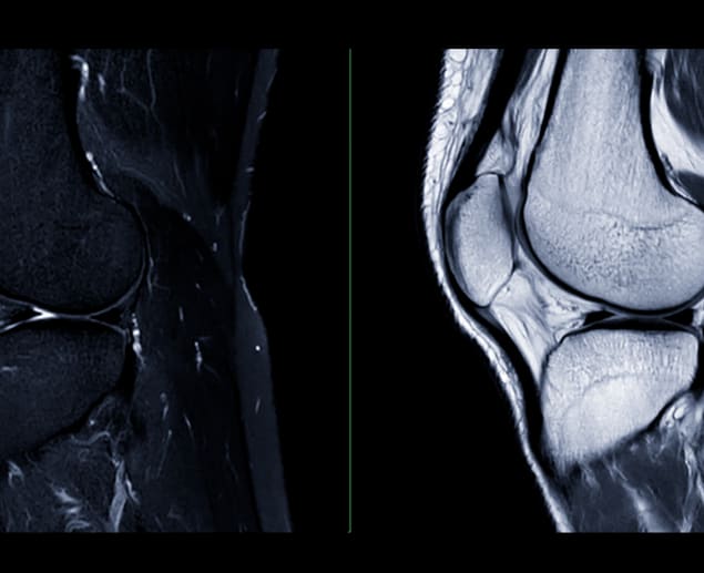
If you’re experiencing knee instability, pain, or swelling, you may be wondering if you’ve sustained an anterior cruciate ligament (ACL) tear. In this article, we’ll explore what an ACL tear is, its common symptoms, and how an ACL tear MRI can help diagnose anterior cruciate ligament injuuries. We’ll also share the causes of ACL tears and what the outlook is for you if you’ve had one.
What is an ACL Tear?
An anterior cruciate ligament tear (ACL) is a common knee injury that can cause knee instability. The anterior cruciate ligament is one of the main ligaments in your knee, and its job is to keep your shinbone from sliding forward and to stabilise your knee when you twist, turn or change direction suddenly or where you jump and land. It’s possible to tear this ligament if you play sports where you might be required to make these movements, such as football, basketball, skiing and tennis, so it's a common injury among athletes.
Knee ligament damage can also occur in the lateral collateral ligament to the side of the knee, as well as the posterior cruciate ligament to the back of the knee and the medial collateral ligament on the inside of the knee.
ACL Tear Symptoms
An ACL tear can cause sudden knee instability and pain, often making it hard to move or bear weight on the injured leg. Here are the common symptoms to watch for if you suspect an ACL injury:
-
Popping sensation: You may feel or even hear an audible ‘pop’ in your knee at the time of injury.
-
Severe pain: Intense knee pain that may prevent you from continuing your activity right after the injury.
-
Rapid swelling: The knee may swell quickly following the injury, usually within a few hours.
-
Loss of range of motion: It may become hard to move or bend your knee during activity fully or when trying to extend or flex the knee when sitting down.
-
Instability or ‘giving way’: The knee might feel unstable, especially during twisting or pivoting movements. Some people with an ACL tear can even find walking challenging because the ACL is so important for stabilising the knee and shinbone.
-
Difficulty bearing weight: You might not be able to put weight on the knee at first, although some people find this improves with time.
-
Feeling uneasy when moving your knee: You may feel apprehensive or cautious when trying to move in ways involving twisting or changing direction.
-
Joint stiffness: You may feel as if your knee is very stiff because of swelling or inflammation., which can further limit your mobility.
-
Other symptoms: ACL tears are often accompanied by other injuries, such as a meniscal tear or damage to the cartilage in the knee, so you may also feel symptoms of these injuries, such as locking or ‘catching’ in the knee
Knowing which symptoms to look out for can help you get a timely MRI scan and diagnosis of ACL injury, which can significantly help your recovery and outcome from an ACL tear.
Will an ACL Tear Show on an MRI?
Magnetic resonance imaging (MRI) is a powerful tool for detecting complete ACL tears, so experts consider it a reliable way of diagnosing the injury. Studies have shown (2022) that MRI has a sensitivity of 93% to 100% in detecting complete tears.
MRI is less effective at detecting partial tears, which can be trickier to diagnose. They often look similar to complete tears on MRI, so your clinician may recommend an advanced MRI technique, such as 3D imaging or angled scans (oblique axial imaging), which can help identify partial tears more clearly. After an MRI, your doctor may compare the images with arthroscopy- a minimally invasive surgical procedure that lets them look directly at the ACL with a tiny camera - for the clearest picture of your injury.
It's important to bear in mind, though, that while MRI is a fantastic tool for spotting ACL injuries, it doesn’t always show every detail, especially with more complicated tears or where there are further injuries to the knee.
The timing of your MRI can be a significant factor in how well it shows your ACL tear. It works best to have the MRI a few days to a couple of weeks after the injury because swelling and bleeding around the tear make it easier to detect. However, if it’s carried out too soon, the same swelling can sometimes hide the damage, making it more challenging for experts to assess the ACL fibres for how severe the tear is. Waiting too long can also make diagnosis tricky since the ligament may start to heal, especially in partial tears, changing how it looks on an MRI.
What Does an ACL Tear Look Like on MRI?
How your ACL tear will look on MRI depends on the timing of your scan and whether it’s a complete or partial tear, but your clinician will look for key signs of each on your MRI results.
The ACL may appear as a bright spot, which is a sign that it’s damaged or inflamed. A complete tear will often show as a gap or ‘space’ in the ligament, and the gap may look wavy.
Swelling and bleeding can create a cloudy appearance on your scan results, sometimes making it difficult to see all the details. Your clinician will look for broken or disconnected ACL fibres to confirm a tear. They may also look for evidence that the ACL is in a position different from usual. For example, if the angle of the ACL has changed by around 15 degrees or more, this suggests it is torn or pulling away from the shinbone or the point where it is attached (tibial insertion).
If the ACL looks bright on certain MRI images and the angle is normal, it’s more likely to be a partial tear rather than a complete one. Unlike a complete tear, where there is a clear gap in the ACL, a partial tear may still show a connection of the ligament fibres, although they may appear thinner or irregular. The shape of the ACL may also look wavy or irregular in form instead of straight and smooth. This can be a sign of damage.
Clinicians will also look for other structural issues or indirect signs that could indicate a torn ACL. For example, anterior tibial translation (ATT) - an abnormal space between the tibia and femur that is common in the case of a complete tear (ACL rupture), or a notch in the lateral femoral condyle which can be caused by the same internal rotation of the knee that can tear the ACL. In fact, the movement and instability of a torn ACL can also cause other tears, such as the posterior horn of the medial meniscus.
Diagnosing an ACL Tear
Physical Examination
A physical examination is an important first step in diagnosing an ACL tear. Your clinician will examine your knee for signs of swelling, and they’ll check your range of motion and how tender your knee is. They’ll also check the joint's stability by asking you to perform a range of movements.
Patient History Assessment
Your doctor will ask you about what you were doing at the time of injury, what symptoms you’ve experienced and whether you’ve had any previous knee injuries.
Lachman Test
The Lachman test is one of the most reliable clinical tests for diagnosing ACL tears. You’ll be asked to lie down, and your doctor will stabilise the upper leg (femur) while pulling the shinbone (tibia) forward. If your shinbone moves forward more than usual, this suggests a more severe injury.
Anterior Drawer Test
Your doctor will flex your knee at 90 degrees while you’re lying down. They will then pull your shinbone forward while stabilising your foot. If the shinbone moves forward more than normal, it’s a strong sign that the ACL has a tear.
Pivot Shift Test
With you lying down, your doctor will push your knee outward (applying ‘valgus stress’) while simultaneously turning your shinbone inward and bending the knee. If they feel a ‘clunk’ while bending the knee, it suggests an injury to the ACL.
MRI Scan
Your doctor will recommend an MRI to provide detailed images of the soft tissues and ligaments around the knee areas, looking for primary and secondary signs of ACL injury. This will help them get a closer look at the injury and check for any associated damage, such as a meniscus tear.
X-Ray
Your doctor may also recommend an X-ray simply to rule out other fractures.
CT Scan (If Necessary)
If your doctor is concerned that your injury is complex or that you have other injuries that an MRI can’t pick up clearly, they may recommend a CT scan to get detailed images of the bony structure of the knee. This can help them to plan the correct surgical treatment.
Arthroscopy (If Necessary)
If the physical tests, MRI and CT scan cannot provide sufficient information for a diagnosis, your clinician may recommend arthroscopy. This minimally invasive surgical procedure allows your doctor to insert a tiny camera into the knee to look directly at the ACL. Experts consider arthroscopy the gold standard for diagnosing ACL injuries and meniscal tears.
Causes of ACL Tears
ACL tears are complicated injuries that can happen for several reasons. Knowing the causes may help you prevent them from happening.
-
Non-contact injuries: Most ACL injuries happen without any direct contact, usually during activities and sports that require sudden turning, jumping or landing. For example, quickly rotating while your foot is planted can cause an ACL tear - this can affect basketball players. Skiers can also be at risk because they may twist their knees while falling, destabilising the joint.
-
Contact injuries: Contact sports, where you collide with other players, such as football, can also cause ACL tears.
-
Biomechanical factors: How you align your knee in shape or position during certain activities can make an injury more likely by putting more stress on the ACL.
-
Fatigue and physical condition: If you’re exhausted during exercise, your control over your movements decreases. Similarly, if you’re out of shape, you may also lack the strength to control your movements. This can affect your knees' stability during exercise, putting extra pressure on the ACL.
-
Anatomical or genetic factors: You may simply have a narrower space in your knee joints, making them less stable and more likely to suffer injury.
-
Previous injuries: Damage to the knee from a past injury can affect movement and stability, putting undue stress on the ACL and increasing the risk of further injury.
Prognosis for an ACL Tear
Recovering from a torn ACL typically takes about six to nine months. However, if you’re a competitive athlete, you might more time for your deficient knee to heal completely before you’re cleared to return to your sport by your sports medicine professional.
Your healthcare provider will let you know when it's okay to start physical activities again. It’s important not to jump back into sports or workouts until you have their approval. Returning too soon can increase your risk of re-injuring your ACL.
Why an MRI Scan is a Good Option
An MRI scan is a great option for diagnosing ACL injuries because it provides detailed images of the soft tissue around the knee, allowing doctors to see the ligament. It is particularly effective for detecting complete ACL tears, with a high accuracy rate of 93% to 100%. MRI can also reveal associated injuries, like meniscus tears, which often occur alongside ACL injuries.
Can MRI Be Wrong About an ACL Tear?
While MRI is a powerful tool for detecting ACL tears, it’s not always 100% correct. Partial tears can be challenging to diagnose with MRI because they appear similar to complex tears.
The timing of the scan is important because swelling and bleeding can mask important injury characteristics while waiting too long can cause the ligament to change in appearance when it starts to heal.
An MRI may not be able to capture the complexity of an injury, especially if other tissues and ligaments are damaged. The quality of the MRI images can also depend on the technique and equipment used, so some scans may not provide a clear view.
How an MRI Scan Detects an ACL Tear
ACL tear MRI scans use strong magnetic fields and radio waves to produce detailed images of the knee, allowing for the detection of any injuries to the anterior cruciate ligament (ACL).
The process begins by aligning the water molecules in the knee using magnets. Radio waves then momentarily disrupt this alignment, and as the molecules return to their original positions, they emit signals that the MRI machine captures.
A computer processes these signals to create detailed, slice-by-slice images, highlighting any areas of damage to the ACL or surrounding structures, such as swelling or tears.
Find an MRI Scan for an ACL Tear
Having a suspected ACL tear can be painful and stressful, but an MRI can provide a clear diagnosis and inform your treatment options. Whether you require rest, pain management, physical therapy or surgery, an MRI offers essential insights for managing your recovery. If you're uncertain about whether an MRI is the right choice for you, our team of expert clinicians is here to help you understand your options and decide on the best steps to take for your knee health and to prevent further injuries.
Sources:
Anterior cruciate ligament (ACL) injury. (2023). https://bestpractice.bmj.com/topics/en-gb/589
Anterior Cruciate Ligament (ACL) Injuries. (2022). https://orthoinfo.aaos.org/en/diseases--conditions/anterior-cruciate-ligament-acl-injuries/
Anterior cruciate ligament injury. (2023). https://www.bupa.co.uk/health-information/knee-pain/anterior-cruciate-ligament-acl-injury
ACL injury. (2022). https://www.mayoclinic.org/diseases-conditions/acl-injury/symptoms-causes/syc-20350738
ACL Tear. (2023). https://my.clevelandclinic.org/health/diseases/16576-acl-tear
Anterior cruciate ligament tear. (2024). https://radiopaedia.org/articles/anterior-cruciate-ligament-tear?lang=gb
Basha, MI. (2022). Comparative study of arthroscopic and MRI findings of injured Knee. https://www.orthopaper.com/archives?year=2022&vol=8&issue=1&ArticleId=3044
Knee ligament surgery. (2021). https://www.nhs.uk/conditions/knee-ligament-surgery/
Perini, JA., et al. (2022). Influence of type I collagen polymorphisms and risk of anterior cruciate ligament rupture in athletes: a case-control study. https://bmcmusculoskeletdisord.biomedcentral.com/articles/10.1186/s12891-022-05105-2


